Seeing Heat: How Thermography Works & What It Can Reveal

CT Thermography • June 24, 2025

Share this article

Thermographic imaging—which may also be referred to as thermal imaging or infrared thermography—allows us to see heat patterns that are typically invisible to the naked eye. It's a noninvasive technique that's becoming increasingly common as a screening tool in the healthcare field. Its usage allows medical professionals to find potential health issues before they become serious problems. In this post, we'll explore how this technology works and why it matters for your health. 


What Is Thermographic Imaging?

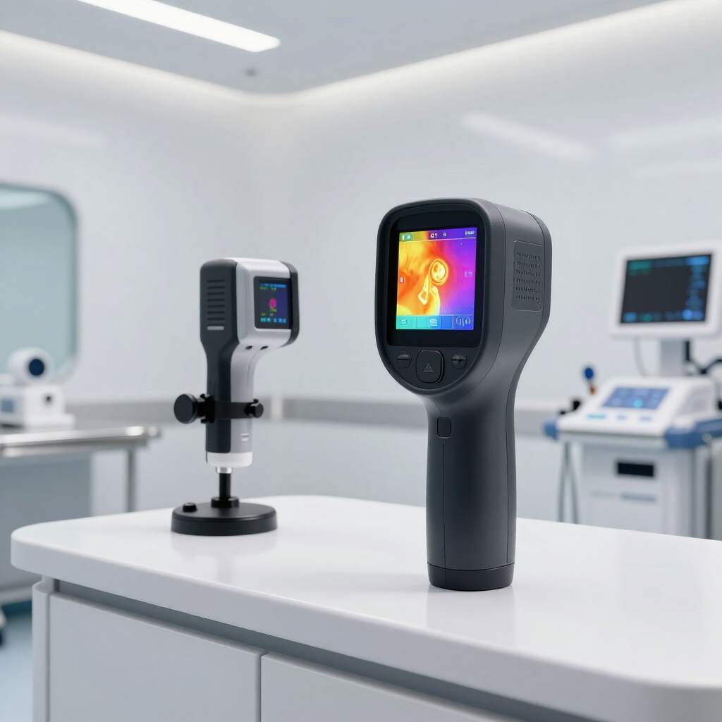
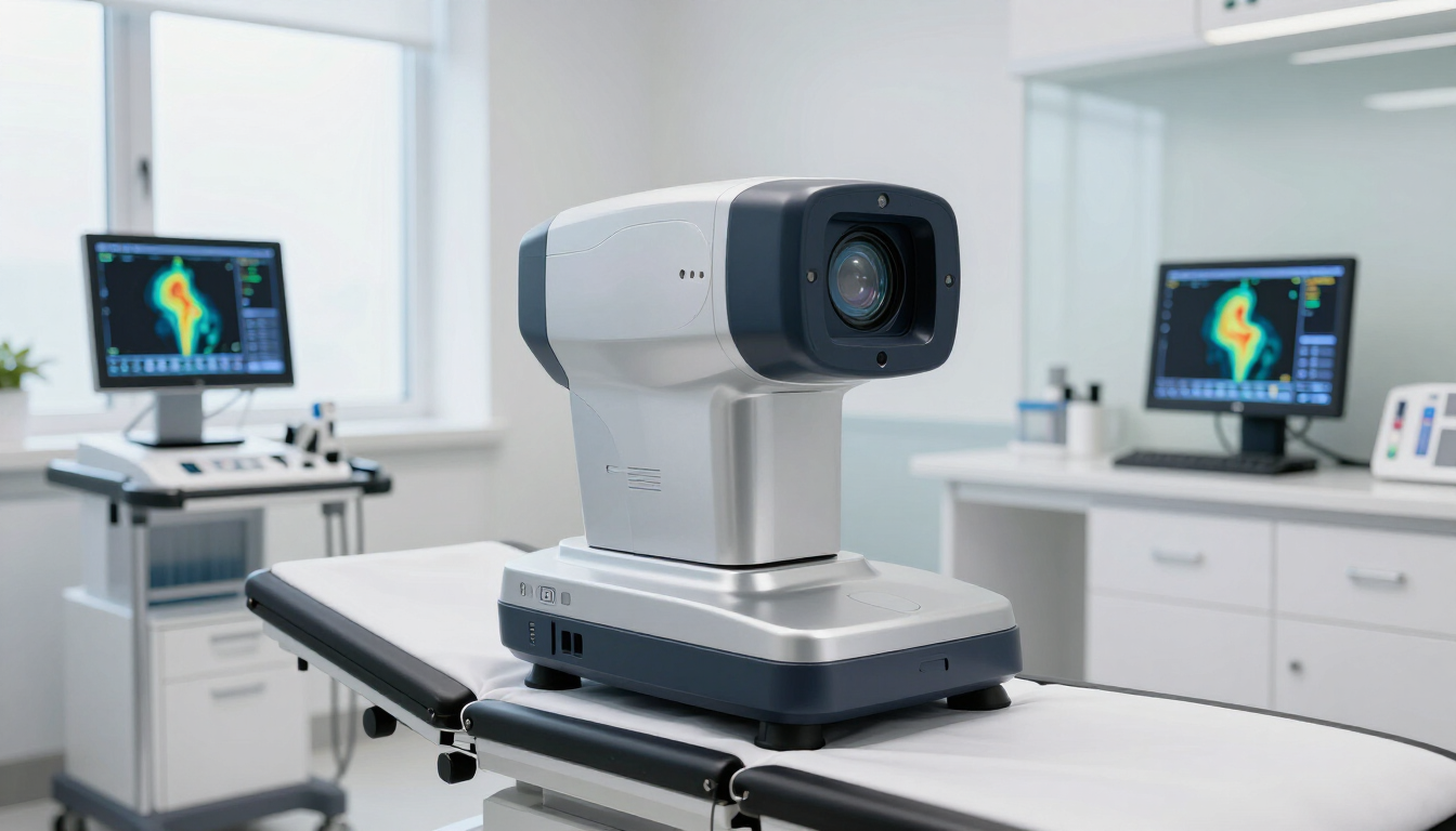
Using special cameras that detect infrared radiation (heat), thermographic imaging provides a unique look at the human body. Rather than capturing light, thermal cameras detect any temperature above absolute zero. 

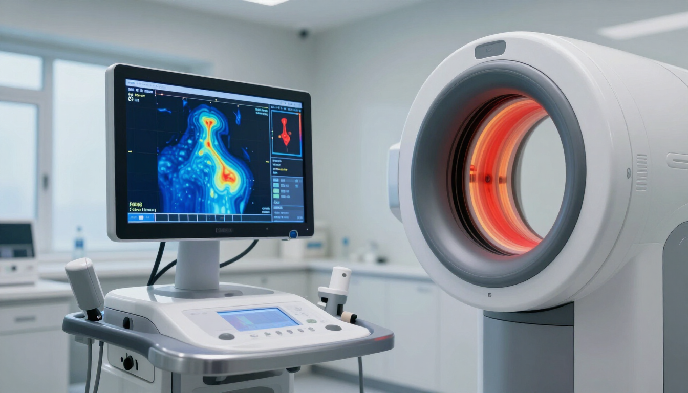
The temperatures are converted to a range of colors to make the visualization meaningful. Warmer areas are bright colors like whites, reds, oranges, and yellows. Cooler areas are darker colors, such as purples, blues, and greens. 

It can be thought of as a heat map of your body. By comparing the patterns of heat in a patient with those of a healthy person, medical professionals can get unique insights into various health conditions. 


How Does Thermographic Screening Work?

Much like a traditional, light-based camera, thermal cameras are relatively simple. Here's how they work:

  • Heat detection : The camera measures the heat coming from your body
  • Temperature measurement : The temperatures are measured with a high degree of precision
  • Image creation : Those temperatures are converted to a color-coded image
  • Pattern analysis : Healthcare professionals examine the patterns for abnormalities

The human body is very symmetrical when it comes to heat patterns. Under normal conditions, the temperature distribution on the right side matches closely with that of the left side. An asymmetrical heat pattern or unusual "hot spot" may indicate a medical condition that warrants further attention. 


The Body's Temperature Story

So, what exactly does the body's temperature tell us about health? Temperature changes often accompany physiological processes:

  • Increased heat : Increased blood flow, caused by inflammation, infection, or increased metabolic activity, often raises heat levels. 
  • Decreased heat : Conversely, a reduction in blood flow often results in decreased heat. This reduction can indicate blockages, nerve damage, or other circulatory issues. 

What makes this exciting for the medical profession is that these changes often occur before other noticeable symptoms do. Thanks to the precision of thermographic imaging cameras, the tests can potentially detect problems before other methods. 


Thermographic Imaging Applications in Health Screening

These insights provided by thermographic imaging are useful for a variety of healthcare diagnosis needs:


Breast Health Assessment

Perhaps the most recognizable use of thermographic imaging is in breast health. Cancerous growths often trigger higher metabolic rates and increased blood supply, resulting in warmer areas in thermal visuals. These differences may become evident before traditional scans detect changes, allowing for earlier treatment and better health outcomes.

Keep in mind, breast thermography isn't a substitute for mammograms. While thermography detects physiological variations, mammograms capture structural anomalies.


Circulatory Issues

Since blood flow is a major aspect of what thermography shows, it's easy to see how beneficial it can be for detecting circulatory issues. These include:

  • Vascular disorders
  • Poor circulation in the extremities
  • Deep vein thrombosis risk
  • Diabetic complications affecting circulation


Inflammatory Conditions

Since inflammation also causes heat, the technology can help monitor inflammatory conditions, including:

  • Arthritis
  • Tendonitis
  • Bursitis
  • Nerve inflammation

The ability to monitor these over time can be a great way to get an objective assessment of how well treatment options are working.


Muscle and Soft Tissue Injuries

If you've ever worked out really hard, you might have found that the muscles being worked feel a little warm. This shows up in thermography. It can help athletes and their healthcare providers:

  • Identify overuse injuries before they cause significant damage
  • Monitor healing progress
  • Guide rehabilitation efforts


Dental Issues

There are also a few dental issues that show heat variations before other symptoms, such as:

  • Hidden infections
  • TMJ (temporomandibular joint) disorders
  • Nerve irritation


Advantages of Thermographic Screening

The increasing use of thermographic imaging is due to several distinct advantages:


Early Detection

One of the clearest benefits is early identification. With thermographic imaging, some issues might be spotted months—or even years—before structural changes become evident using other diagnostics.


Completely Non-invasive

Some screening methods are highly invasive. Although they are essential for confirming a diagnosis or detecting problems, repeating them frequently can cause problems. Thermography doesn't have this problem, as it:

  • Involves no radiation
  • Requires no contact with the body
  • Causes no pain or discomfort
  • Needs no injections or contrast agents


Whole-Body Assessment

Most diagnostic methods target a specific body part. Thermographic imaging provides a full-body overview, revealing various possible concerns in one session.


Limitations of Thermographic Imaging

Despite its many benefits, thermographic imaging has its limitations:

  • It doesn't provide structural data like X-rays, CT scans, mammograms, or MRIs do
  • It can indicate that something might be wrong, but it does not give a complete diagnosis on its own
  • Effects can be impacted by environmental factors or recent physical activity
  • The quality of interpretation from results varies according to the practitioner's experience

For these reasons, thermography is an additional tool in the fight to detect, prevent, and treat illness—but shouldn't be considered the only tool. 


What to Expect During a Thermographic Scan

If your healthcare provider has suggested a thermographic scan, here's what you can expect during the process:

  • You'll be asked to sit in the room for 15-20 minutes to acclimate to the room temperature and avoid outside temperatures that could affect the results.
  • The technician will position you in the best way to obtain the necessary thermal images. The specialized camera doesn't touch your body.
  • After the images are taken, a trained thermographer interprets them, looking for specific patterns or abnormalities.

The entire process takes around 30-60 minutes.


Thermographic Imaging: A Preventative Health Resource

Thermographic imaging offers healthcare providers a unique look at how your body functions. In doing so, it can potentially reveal issues that otherwise could not be detected as early. Its ability to detect irregularities that aren’t always visible using other tools adds meaningful depth to diagnostics.

While it shouldn't be viewed as a replacement for other medical tests, it provides complementary information that can help better guide healthcare decisions. If you or your healthcare provider thinks you can benefit from thermography, CT Thermography can help. Contact us today for more information.

Recent Posts

Peaceful late summer sunlight in New England representing wellness.
August 10, 2026
Learn how to prepare for your late-summer thermography appointment. Get essential tips on sun exposure, tanning, and timing your screening for accurate results.
A private medical imaging room for thermography in Westport, CT.
August 6, 2026
A comprehensive guide on what to expect during your thermography appointment in Westport, CT. Learn about our private, temperature-controlled imaging process.
Modern thermography clinic suite in Connecticut
July 27, 2026
Discover how adjunctive thermal imaging enhances integrative care. Learn about PACT-certified screenings and physician-led reporting for Connecticut practitioners.
Professional thermography imaging equipment on a tripod in a clinic.
July 20, 2026
Explore the different thermography imaging options including breast, head and neck, upper body, and full body screenings to determine which best fits your wellness goals.
A clean and professional thermography imaging room at CT Thermography.
July 13, 2026
A comprehensive guide to full body thermography for men. Learn about the non-invasive imaging scope—from head to testicular health—and the appointment flow at CT Thermography.

A patient reviewing their thermography report on a laptop computer.
July 6, 2026
Learn what to expect in your thermography report, from clinical impressions to secure portal delivery. This guide helps you prepare for discussions with your provider.
Professional medical thermography imaging setup with infrared camera and monitors.
June 22, 2026
Learn how CT Thermography uses a three-level interpretation process by board-certified thermologists and FDA 510(k)-cleared equipment for quality results.
Modern and professional thermography imaging room.
June 8, 2026
Learn why a baseline scan and comparison imaging are essential in medical thermography for monitoring stable thermal patterns and proactive wellness.
A private, professional thermography screening room at CT Thermography.
May 4, 2026
Curious about your first breast thermography appointment? Learn about the private, contact-free process, imaging angles, and how you receive your wellness report.
A calm person preparing for a wellness screening with a glass of water.
April 6, 2026
Follow this professional thermography preparation checklist to ensure accurate imaging results. Learn why skin temperature and hydration matter for your screening protocol.
Show More